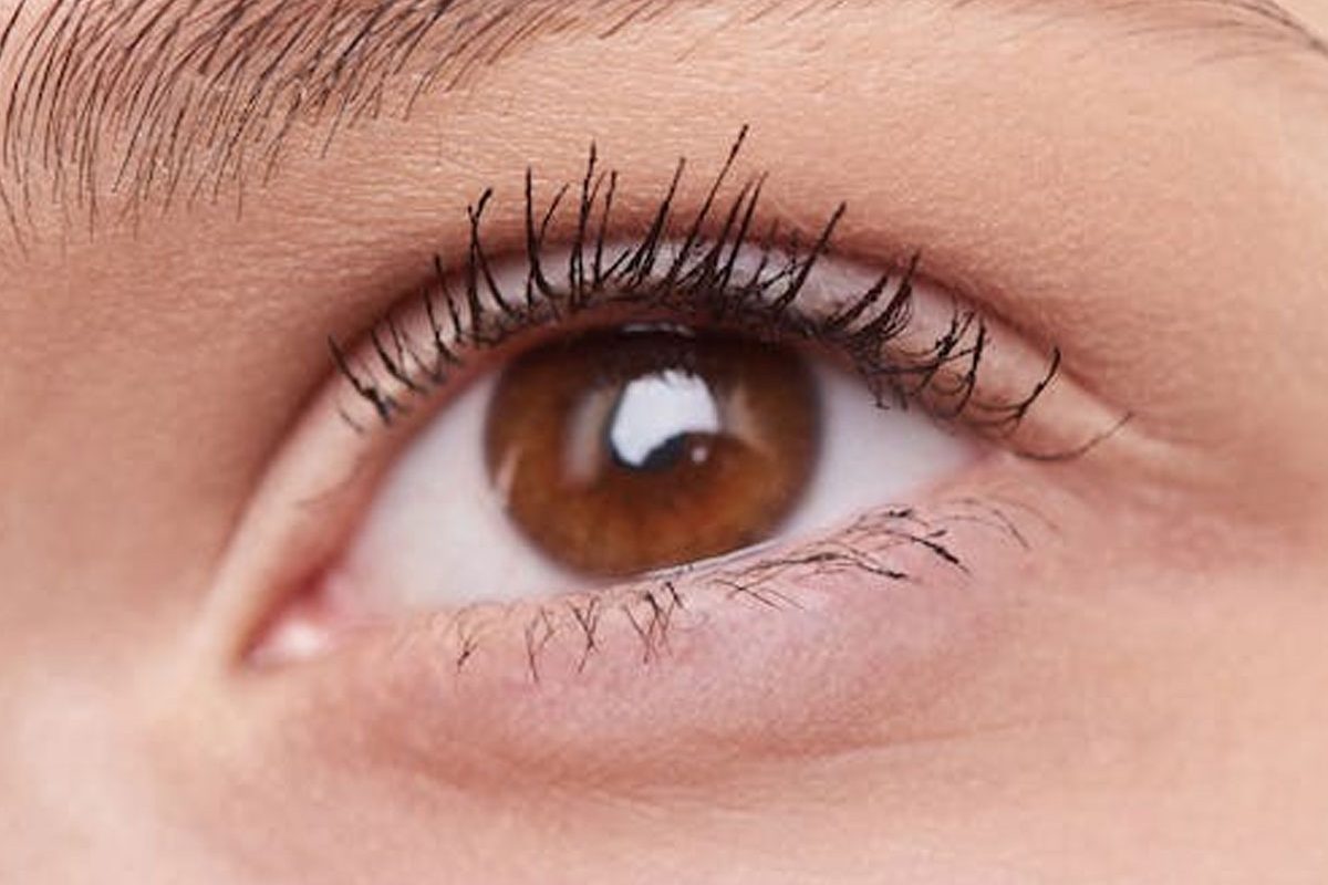

If you’ve been a spectacle-wearer for any length of time, the idea of contact lenses can be appealing. Unlike glasses, contacts don’t fog up, become spattered with rain, get knocked off your face during sport, or slide down your nose. With modern advancements in contact lens materials and design, many Canberra patients find that they can be very successful with contact lens wear.
Am I suitable for contact lenses?
Determining whether contact lenses are right for you – and which ones – can only be done with an optometrist. During your appointment, your optometrist will assess factors such as:
- Your prescription and vision. Contact lens manufacturers produce a limited, though extensive, number of prescriptions.
- The health of the surface of your eye, which is where the contact lens will sit.
- Signs and symptoms of dry eye. Dry eyes can impact your success with contact lens wear, so this will need to be properly managed first.
You will also have a discussion with your optometrist about your expectations with contact lenses, such as how often you intend to wear them and for what sort of activities. This will help your optometrist select the right type of lens for you to trial first.
Are contacts good for my vision?
Many patients find their vision in contacts is just as good as what they can achieve in their glasses. There can be occasions where you may find your sight in glasses is slightly crisper than in contacts, such as if you have astigmatism or have been fitted in a multifocal contact lens. However, lens designs nowadays still allow for very clear, functional vision even for these patients.
Contact lens wear is associated with an increased risk of adverse events, such as eye infections. However, by following your optometrist’s instructions regarding wearing habits, lens hygiene, and regular contact lens aftercare appointments, you can greatly reduce your risk.
If you’re interested in how contact lenses can benefit you, your vision, and your lifestyle, call Junic Eye Care (Eyecare Plus Coombs) today on (02) 6152 8585.
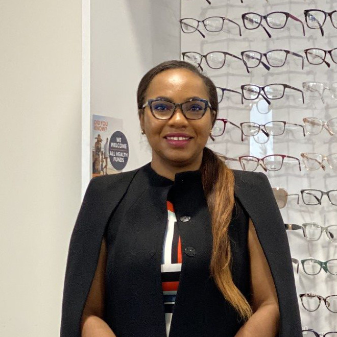
CANBERRA OPTOMETRIST
Juliet obtained her Doctor of Optometry degree from the University of Benin, Nigeria in 2006. She completed an internship programme before migrating to Australia, where she completed a master’s degree in public health at the University of Sydney in 2014. Following this, Juliet obtained a Master of Orthoptics from the University of Technology Sydney (UTS) in 2017.
Juliet has completed her competency in optometry examination with OCANZ (Optometry Council of Australia and New Zealand), and obtained her ophthalmic prescribing rights from ACO (Australian College Of Optometry Victoria). Juliet has worked in various positions, including retail Optometry, the Ophthalmology Department at Canberra Hospital, and more recently, at the John Curtin School of Medical Research (ANU).
As a dedicated Canberra optometrist, Juliet is passionate about helping people with low vision, and binocular vision anomalies hence her interests in Low Vision Rehabilitation, Eccentric Viewing Training and Paediatric optometry.


")

")


")