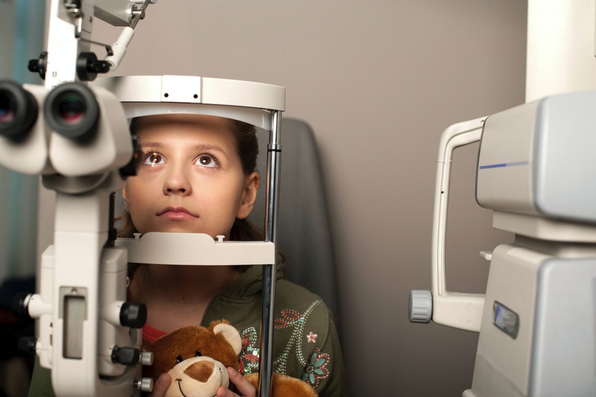
BOOK ONLINE
(02) 6152 8585
Toll Free : 1 123 456 78910

Category Archives: Blog

Headaches, Migraines, and My Eyes

Canberra optometrists regularly encounter patients reporting concerns with headaches. Considering that your eyes are housed within your head, headaches are often experienced around the eye area and can often come on after reading or other visual tasks, this is no surprise. However, the relationship between eyes and headaches or migraines can often be a little more complex than one might expect.

Headaches cause vision issues.

There are different types of headaches. Understanding and managing headaches and migraines can be a special area of interest for a number of healthcare professionals, such as neurologists or physiotherapists.

The most common headaches that are associated with visual disturbances are migraines and cluster headaches.1

  • Migraines: These are intense headaches that are often associated with other symptoms such as general malaise, vomiting, and hypersensitivity to stimulation such as lights or noise.1,2 Visual symptoms of a migraine may precede the actual head pain – this is termed a visual aura and is often described as the sensation of tunnel vision, seeing zigzagging, shimmering, or flashing coloured lights,1 or like looking through a kaleidoscope. Your vision may also become blurry. Some cases of migraine, called a visual migraine or retinal migraine, present as only the visual symptoms without the headache.
  • Cluster headache: As the name suggests, cluster headaches occur in clusters, for example, every day for several months, then disappear for months or years before the next cluster. Ocular and visual symptoms of cluster headaches can include watery, red eyes, drooping eyelids, and changes to pupil size.1

Vision issues can cause headaches.

Studies have shown that uncorrected refractive error, that is, not wearing the right (or any) glasses or contacts when you need them can be associated with headache,3 mainly through eyestrain. The link between uncorrected refractive error and migraine is a little more controversial but there is some suggestion that there is an association with uncorrected astigmatism and differences in prescription between the eyes.4

Risk factors for headaches due to refractive error include3

  • Prolonged screen-time
  • Having more than one type of refractive error
  • Moderate long-sightedness
  • Astigmatism

Even if you don’t typically need glasses, eyestrain can also arise from overusing your eyes, such as excessive reading, screen-time, or focusing on close detail for too long without a break. This causes the muscles of your eyes to work in overtime, which results in fatigue, strain, and headache. Eyestrain headaches are often felt around the temples or the brow.

In addition to eyestrain, certain eye diseases are associated with headache as a symptom. These include:1

  • Acute angle closure glaucoma, involving a sudden spiking of the pressure inside your eye.
  • Ocular ischaemic syndrome, which occurs due to chronic poor blood flow to the eye.

Other things entirely can cause both headaches and vision issues.

There are a number of conditions not directly related to the eyes that can result in both visual symptoms and headaches. These include:

  • Temporal arteritis. This is a medical emergency involving inflammation of the arteries around your temple. You may notice a deterioration to your vision and throbbing ache on one side of the head.
  • Intracranial hypertension. Also known as pseudotumor cerebri, this refers to raised pressure inside the skull, which can result in a dull headache at the back of the head, worse at night or in the morning on waking. Visual symptoms include blurring and double vision.5

If you are experiencing unusual headaches, it’s always worth a visit to your Canberra optometrist to rule out visual or ocular causes, even if you don’t think they’re related. Call Junic Eye Care today on 02 6152 8585 to book your appointment.

References

  1. Verywell Health. Headaches and Your Vision. https://www.verywellhealth.com/. 2022. Available at: https://www.verywellhealth.com/vision-and-headache-3422017. (Accessed February 2023).
  2. Healthdirect. Migraine. https://www.healthdirect.gov.au/. 2021. Available at: https://www.healthdirect.gov.au/migraine#symptoms. (Accessed February 2023).

Sudden Flashes and Floaters – Should I Find an Optometrist Near Me Immediately?

For some people, the appearance of sudden flashing lights in the vision accompanied with floating specks or lines can be alarming. They understandably ask the question “Should I find an optometrist near me immediately?”

For others, it may be an odd observation that is soon forgotten amidst the general busyness of everyday life.

So, what causes flashes and floaters? And more importantly, what are you supposed to do about them?

What are Flashes and Floaters?

Also known as photopsia, these flashes of light are often described as a lightning arc noticed out of the corner of your eye, often more noticeable when your environment is dim. They can also be described as seeing a reflection off a surface.

Floaters are often mistaken for a fly or other insect in your vision. They can also present like squiggles or be cobweb-like in appearance. You may see one or a few discrete floaters, or you may experience a sudden shower of many tiny dots.

What Do They Mean?

Flashes and floaters originate from inside the eyeball – there is actually no lightning in the distance, nor is there a fly buzzing around your face. There are two common reasons for flashes and floaters.

Posterior vitreous detachment (PVD)

A PVD is a normal age-related change to the vitreous gel occupying the back half of the eyeball. As we age, this gel loses its solid molecular arrangement and slowly begins to liquify. Because the vitreous is attached to certain parts of the light-sensing retina, as it collapses in on itself, it can tug at these points. This tugging mechanically stimulates the perception of light from the retina, which is what you see as photopsia. Floaters during a PVD can occur from strands of vitreous floating around pockets of liquid, or from tufts of retinal tissue that have been pulled off as the vitreous shrinks away from its points of attachment.

Retinal detachment.

A retinal detachment is considered a sight-threatening eye condition. The retina lines the inside of the eyeball. During a detachment, it peels away from its underlying tissues, much like wallpaper peeling away from the wall. The retina relies on support from the underlying tissues to function – if it becomes detached, it is no longer able to receive blood supply or nutrients, nor is it able to send on its neural signals for vision. Flashes during a retinal detachment are due to mechanical stimulation of the sensory photoreceptors, while floaters can be from fragments of retinal tissue or blood cells from broken retinal capillaries. A retinal detachment may only involve a small area of retina or can be extensive; it may involve the macula (affecting your central vision), or the macula may be preserved. Another telling symptom of retinal detachment includes noticing a shadow or dark curtain across part of your sight.

What Should I Do?

Any observation of flashes with or without floaters should be investigated by an optometrist promptly. Your local optometrist is well-equipped to assess the state of your retina and advise whether it’s a normal PVD that requires monitoring or a retinal detachment that requires urgent referral to an ophthalmologist.

You can expect to have eyedrops that will dilate your pupil so your optometrist can get a good view of the retina. With these drops in effect, you will not be able to drive for a couple of hours afterward, you’ll be very glare sensitive, and your near vision may be blurry.

The sudden onset of flashes and floaters should not be ignored, even if your vision is otherwise fine. If you experience these symptoms, contact your local Canberra optometrist on the same day for further advice on what to do.

For all appointments visit www.juniceyecare.com.au or call (02) 6152 8585 and book in with Canberra Optometrist.

Will Contact Lenses Help My Vision?

If you’ve been a spectacle-wearer for any length of time, the idea of contact lenses can be appealing. Unlike glasses, contacts don’t fog up, become spattered with rain, get knocked off your face during sport, or slide down your nose. With modern advancements in contact lens materials and design, many Canberra patients find that they can be very successful with contact lens wear.

Am I suitable for contact lenses?

Determining whether contact lenses are right for you – and which ones – can only be done with an optometrist. During your appointment, your optometrist will assess factors such as:

  • Your prescription and vision. Contact lens manufacturers produce a limited, though extensive, number of prescriptions.
  • The health of the surface of your eye, which is where the contact lens will sit.
  • Signs and symptoms of dry eye. Dry eyes can impact your success with contact lens wear, so this will need to be properly managed first.

You will also have a discussion with your optometrist about your expectations with contact lenses, such as how often you intend to wear them and for what sort of activities. This will help your optometrist select the right type of lens for you to trial first.

Are contacts good for my vision?

Many patients find their vision in contacts is just as good as what they can achieve in their glasses. There can be occasions where you may find your sight in glasses is slightly crisper than in contacts, such as if you have astigmatism or have been fitted in a multifocal contact lens. However, lens designs nowadays still allow for very clear, functional vision even for these patients.

Contact lens wear is associated with an increased risk of adverse events, such as eye infections. However, by following your optometrist’s instructions regarding wearing habits, lens hygiene, and regular contact lens aftercare appointments, you can greatly reduce your risk.

If you’re interested in how contact lenses can benefit you, your vision, and your lifestyle, call Junic Eye Care (Eyecare Plus Coombs) today on (02) 6152 8585.

When Should You See an Optometrist and What Happens During an Eye Test?

In Australia, “optometrists play a key role in preventative care, early detection and treatment of eye and vision problems, and detection and referral of systemic conditions that affect the eye.” Much like GPs are to the medical profession, optometrists are often your first point of call when you have an eye or vision concern.

Should I see an optometrist regularly?

To maximise your chances of maintaining good vision for life, it’s important to have routine eye tests. Even if you feel your sight is perfect and you have no issues, many eye diseases are asymptomatic in their early stages. It can also be difficult to know whether your vision is considered normal or not if you’ve become used to the quality of your sight over time.

If your eyes and vision are known to be healthy, the current recommendation by most optometrists for a regular eye test is:

  • Every 2 years if you are under the age of 65 years old.
  • Every year if you are 65 years or older.

If you do have a condition that needs closer observation, your optometrist will advise you how frequently you should be checked. Outside of your routine visits, if you experience any worrisome symptoms such as eye pain or decreasing vision, your optometrist should be your first stop.

What happens during an eye test?

A comprehensive examination is often tailored to the individual – such as using age-appropriate tests or additional testing relevant to a known concern. A normal routine check-up includes:

  • Reading letters on a screen to test visual acuity
  • Examining the appearance and health of the front and back of the eyeball
  • Measuring the pressure inside the eye

In addition to these, your optometrist may regularly do other tests such as checking your pupil reactions or taking retinal photos and other scans.

At Junic Eye Care, we provide comprehensive eye tests for patients of all ages. If you’re overdue for your routine visit, or if you have any eye or vision-related concerns at all, book your appointment now on 02 6152 8585.

References

Sustainability for optometry and primary eye health care. https://treasury.gov.au/sites/default/files/2019-03/360985-Optometry-Australia.pdf

2022 Vision Index.

https://www.optometry.org.au/wp-content/uploads/GVFL/Vision_Index/2022-Vision-Index-Report.pdf

Can Sleep Apnea Affect Your Eyes?

Obstructive sleep apnoea (OSA) is a common sleep disorder characterized by repetitive episodes of partial or complete upper airway obstruction associated with hypoxemia and re-oxygenation sequences. OSA is more prevalent in men than women. It increases 2-3 times in persons more than 65 years old but sometimes can be seen in children with Adenotonsillar hypertrophy. The most important risk factor is a 10% weight gain which increases the risk of developing OSA by six times.

Sleep apnoea has various ophthalmic presentations which includes:

Floppy Eyelid Syndrome (FES):

This is a condition where the upper eyelid becomes elastic and easily folded upwards due to mechanical trauma to the eyelids during sleep. This is usually seen in association with papillary conjunctivitis, eyelid trauma and corneal epithelial erosions. Seen commonly in obese patients due to weak tarsus.

Glaucoma:

Glaucoma is an optic neuropathy or injury to the optic nerve which manifests as visual field defects. Glaucoma occurs in OSA patients because of vascular and mechanical factors.  Vascular factors include periods of hypoxia followed by oxidative stress during reperfusion. Mechanical factors comprise increased IOP at night due to changes in sleep architecture and increased sympathetic tone.

Nonarteritic Anterior Ischemic Optic Neuropathy (NAION):

NAION is a condition characterized by sudden and painless unilateral vision loss, oedema of the optic disk, and a relative afferent pupillary defect. Patients with OSA are more susceptible to developing NAION because of a combination of hypoxia, oxidative stress and increases intracranial pressure during their apnoeic episodes. Patients with OSA have 16% more probability to develop NAION compared to patients without OSA, and the prevalence of OSA in patients with NAION may be as high as 71%-89%.

Papilledema:

Papilledema is a bilateral swelling of the optic disc. This condition when seen in OSA patients could be because of increase in their intracranial pressure during sleep occurring from hypoxemia during apnea episodes.

Keratoconus:

This is a bilateral condition characterized by progressive thinning of the cornea, irregular astigmatism and protrusion of the central cornea creating conical appearance. Its pathogenesis in patients with OSA is not well understood yet. But males with keratoconus with a family history of OSA is more likely to develop OSA.

Central Serous Chorioretinopathy (CSCR):

This is a serious detachment of the neurosensory retina at the macula. Approximately two-thirds of patients with CSCR have OSA. One theory for this increased prevalence is the presence of augmented oxidative stress, which can produce endothelial cell damage and vasoconstriction.

Why should I get an eye examination if I have sleep apnoea?

Patients with OSA bear an increased risk for several vision threatening ocular conditions. Optometrists as a primary eye care providers are optimally equipped to identify these ocular manifestations. At JUNIC EYECARE PLUS COOMBS we collaborate with ophthalmologists, your primary physician and sleep specialists to appropriately manage these conditions if they arise. These ocular conditions will require close monitoring to prevent permanent vision loss in OSA patients.

What are you waiting for? If you are in Canberra, get an eye checkup at Junic Eyecare Plus Coombs.

www.juniceyecare.com.au

Phone: 02 6152 8585

REFERENCES:

  1. Wong, B., & Fraser, C. (2019). Obstructive Sleep Apnea in Neuro-Ophthalmology. Journal of Neuro-Ophthalmology, 39, 370-379.
  2. Huon, L.-K., Liu, S. Y.-C., Camacho, M., & Guilleminault, C. (2016). The association between ophthalmologic diseases and obstructive sleep apnea: a systematic review and meta-analysis. Sleep Breathing Physiology and Disorders, 1-10.
  3. Skorin, L., & Knutson, R. (2016). Ophthalmic Diseases in Patients with Obstructive Sleep Apnea. The Journal of the American Osteopathic Association, 116, 522-529.
  4. West, S. D., & Turnbull, C. (2016). Eye disorders associated with obstructive sleep apnoea. Curr Opin Pulm Med, 22, 595-601.